Interesting Things with JC #1734: "Tattoo Ink and Your Lymph Nodes"
Tattoo ink doesn't always stay in your skin. Your immune system carries some of it into nearby lymph nodes, where the pigment can remain for decades as immune cells continually pass it from one cell to the next.
Curriculum - Episode Anchor
Episode Title: Tattoo Ink and Your Lymph Nodes
Episode Number: 1734
Host: JC
Audience: Grades 9–12, introductory college, homeschool, and lifelong learners
Subject Area: Biology, immunology, anatomy and physiology, health science, and scientific literacy
Recommended Lesson Length: 60–75 minutes
Instructional Focus: Body-system interaction, immune-cell function, evidence evaluation, and correlation versus causation
Lesson Overview
Learning Objectives:
Trace the movement of tattoo pigment from the dermis through lymphatic drainage to regional lymph nodes.
Explain how macrophage uptake and pigment capture–release–recapture can contribute to tattoo persistence.
Distinguish findings from human tissue studies, animal experiments, case reports, and epidemiological research.
Evaluate health claims by separating established evidence, preliminary findings, statistical associations, and causal conclusions.
Essential Question: How does the immune system respond to tattoo pigment, and what can current evidence legitimately tell us about long-term effects?
Success Criteria:
I can construct and label a model connecting the dermis, lymphatic vessels, macrophages, and regional lymph nodes.
I can explain tattoo persistence using the capture–release–recapture model.
I can identify whether a finding came from human tissue, an animal model, a clinical case, or an observational population study.
I can explain why an association does not by itself establish causation.
I can communicate a conclusion that includes both evidence and limitations.
Student Relevance Statement: Tattoos are common forms of personal expression, but their pigments become part of a biological system. Understanding what happens to that pigment helps students make sense of immune responses, medical imaging, scientific uncertainty, and health information encountered online.
Real-World Connection: Physicians, pathologists, radiologists, researchers, tattoo professionals, and patients may need to understand that tattoo pigment can reach lymph nodes and occasionally complicate the interpretation of pigmented or abnormal-looking tissue.
Workforce Reality: Accurate health-science work requires disciplined observation, careful terminology, evidence classification, and professional judgment. A visually unusual lymph node cannot be diagnosed by appearance alone, and preliminary findings cannot be presented as settled human-health conclusions.
Key Vocabulary
Dermis(DUR-mis): The layer of skin beneath the epidermis into which permanent tattoo pigment is deposited.
Pigment(PIG-ment): A colored material made of particles that absorb or reflect particular wavelengths of light.
Macrophage(MAK-roh-fayj): A white blood cell that engulfs particles, damaged cells, and microorganisms.
Phagocytosis(fag-oh-sy-TOH-sis): The cellular process of surrounding and engulfing particles or other material.
Lymphatic system(lim-FAT-ik SIS-tum): A network of vessels, tissues, and organs involved in fluid balance, immune surveillance, and the transport of lymph.
Lymph node(limf nohd): A small immune organ that filters lymph and provides a location where immune cells encounter transported material.
Draining lymph node(DRAY-ning limf nohd): A lymph node that receives lymph from a particular region of the body.
Inflammation(in-fluh-MAY-shun): A coordinated biological response to injury, irritation, infection, or foreign material.
Lymphoma(lim-FOH-muh): A group of cancers that begin in cells of the lymphatic and immune systems.
Correlation and causation(kor-uh-LAY-shun; kaw-ZAY-shun): Correlation is a statistical relationship between variables; causation means that one factor produces or contributes directly to an outcome.
Narrative Core
Open: A tattoo appears to remain exactly where it was placed, but some of its pigment can leave the skin and accumulate in lymph nodes that drain the tattooed area.
Info: Tattoo needles deposit pigment in the dermis. Immune cells respond to this foreign material, and pigment particles may enter lymphatic drainage either as free particles or after uptake by phagocytic cells. Human tissue research has identified matching tattoo pigments in skin and regional lymph nodes.
Details: Macrophages retain much of the pigment that remains in tattooed skin. Mouse research supports a capture–release–recapture model in which dying pigment-filled macrophages release particles that are engulfed by incoming macrophages, helping the visible tattoo persist without requiring the original cells to survive for a lifetime. Whether every part of this mouse model operates identically in humans remains under investigation.
A 2025 study found prolonged inflammation and altered vaccine responses in tattoo-draining lymph nodes in mice. The effect depended on vaccine type and injection location, so the results do not establish a general reduction in vaccine effectiveness in tattooed people.
A Swedish case-control study reported an association between tattoo exposure and malignant lymphoma. Because it was observational, it could not demonstrate that tattoos caused lymphoma, and the authors called for additional epidemiological research.
Reflection: The central scientific skill is not deciding that tattoos are harmless or dangerous. It is determining what each study measured, what population or model it examined, what conclusion the design permits, and what remains uncertain.
Closing: These are interesting things, with JC.
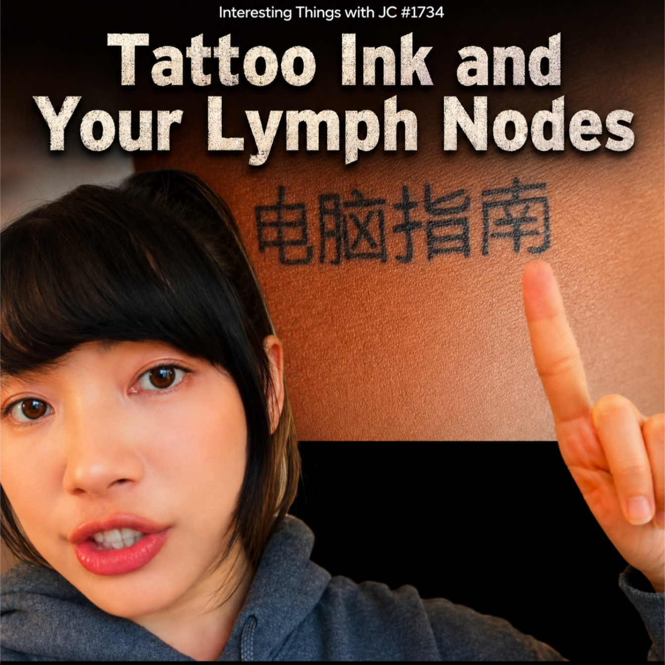
Promotional graphic for Interesting Things with JC #1734: "Tattoo Ink and Your Lymph Nodes." A close-up of a person in a gray hoodie points toward a black Chinese-character tattoo on an upper arm. Large white title text reads "Tattoo Ink and Your Lymph Nodes," with the episode number above it. The image introduces a discussion about how tattoo pigment can migrate from the skin into nearby lymph nodes through the immune system.
Transcript
Interesting Things with JC #1734:
"Tattoo Ink and Your Lymph Nodes"
If you have a tattoo, there's a good chance part of it isn't where the artist put it anymore.
It's inside your lymph nodes.
Doctors have opened patients during surgery expecting to find ordinary lymph nodes and instead discovered some stained black, blue, green, and even red. At first glance, those darkened nodes can look alarming, sometimes even resembling melanoma that has spread.
The explanation is surprisingly simple.
The moment a tattoo is applied, your immune system gets to work. Specialized white blood cells called macrophages recognize the tiny pigment particles as foreign material and begin swallowing them. Many remain in your skin, but some carry that pigment through your lymphatic system into nearby lymph nodes, where it can remain for decades.
For most people, that's considered a normal consequence of having a tattoo. Your lymph nodes are filters. They're designed to trap foreign material before it can travel elsewhere in the body. In other words, they're doing exactly what they're supposed to do.
Researchers have also solved one of the biggest mysteries about tattoos: why they last. The pigment doesn't stay locked inside the same cells forever. As pigment-filled macrophages die, new macrophages arrive and consume the released ink. That continuous handoff helps keep the tattoo visible for years, even though the immune cells themselves are constantly being replaced.
But the ink doesn't simply remain inside those lymph nodes. Researchers have found that it appears to continue interacting with the immune system, and in animal studies, some of that activity can persist for months.
Early laboratory and animal studies also suggest tattoo pigment may alter the behavior of certain immune cells and could reduce antibody responses to some mRNA vaccines when the shot is given in the same area of the body as the tattoo. Those findings appear to be limited to the local lymph nodes that drain that area, and they have not been confirmed as a general effect in people.
There's another question scientists are investigating. A large nationwide study from Sweden found a statistical association between tattoos and lymphoma, a cancer of the lymphatic system, but it did not show that tattoos cause the disease. More research is needed before any conclusions can be drawn.
These are interesting things, with JC.
Student Worksheet
Directions: Listen to the complete episode once without pausing. During the second listening or transcript review, record evidence, study type, and limitations. Answer in complete sentences unless a diagram is requested.
Comprehension Questions:
Into which layer of the skin is permanent tattoo pigment placed?
What immune cells engulf tattoo pigment?
What is the normal function of a lymph node?
Describe the capture–release–recapture process proposed to explain tattoo persistence.
Why might tattoo pigment in a lymph node concern a surgeon or pathologist at first glance?
What two areas of ongoing research are discussed near the end of the episode?
Analysis Questions:
Draw a four-stage model showing tattoo pigment moving from the tattoo site to a regional lymph node. Label the dermis, pigment, lymphatic vessel, and draining lymph node. Add macrophages where supported.
Classify each finding as human tissue evidence, animal experimental evidence, clinical case evidence, or observational population evidence:
Matching pigment identified in tattooed skin and regional lymph nodes
Altered antibody response after vaccination in tattooed mice
A dark lymph node discovered during surgery
A statistical association between tattoos and lymphoma in Sweden
Explain why the statement “tattoos cause lymphoma” goes beyond the evidence described in the episode.
Evaluate the claim “tattoos weaken vaccines.” Identify one supporting observation, two limitations, and a more accurate replacement statement.
The transcript states that some macrophages carry pigment into nearby lymph nodes. Research also permits passive movement of particles through lymphatic fluid. Explain why distinguishing a confirmed destination from an incompletely resolved transport mechanism matters.
Reflection Prompt: In five to seven sentences, explain how this topic changes or strengthens your understanding of the immune system as an active, continuously changing system.
Difficulty Scaling:
Foundation: Complete Questions 1–7 using the transcript and vocabulary list. Use a teacher-provided model template.
On-Level: Complete all questions and include one quoted or paraphrased piece of episode evidence in Questions 9 and 10.
Advanced: Complete all questions and compare the strength of two different study designs.
College Bridge: Complete all questions and add a 150-word evidence appraisal that addresses sample, model, variable, limitation, and permitted conclusion.
Student Output: Submit one labeled biological model, six comprehension responses, five analysis responses, and one reflection paragraph. Scientific terms must be used accurately.
Academic Integrity Guidance: Separate information taken from the episode or assigned research from your own interpretation. Paraphrase accurately, identify the source by author or episode title, and do not present animal findings as confirmed human outcomes.
Teacher Guide
Quick Start:
Begin immediately by playing the complete podcast episode.
Ask students to listen without writing and identify the episode’s central surprise.
Play the episode a second time or distribute the transcript.
Complete the biological-model task before moving into health-claim analysis.
Pacing Guide—Audio First:
0–4 minutes: Play the entire episode; students listen without taking notes.
4–8 minutes: Bell ringer and rapid recall.
8–13 minutes: Replay the episode or conduct a guided transcript read.
13–23 minutes: Teach vocabulary and construct the pigment-pathway model.
23–38 minutes: Complete worksheet comprehension and evidence-classification tasks.
38–50 minutes: Discuss animal evidence, epidemiological evidence, and causal limits.
50–60 minutes: Complete the quiz and exit ticket.
60–75 minutes: Optional assessment planning, source comparison, or extension.
Bell Ringer: Immediately after the first listening, students write: “What detail was most surprising, and what biological question did it create?” Require two sentences.
Audio Guidance: During the first listening, students should focus on the narrative sequence. During the second listening, they should mark each point where the episode shifts from established evidence to preliminary findings or uncertainty.
Audio Fallback: Read the transcript aloud verbatim, assign alternating paragraphs to students, or provide five minutes for silent reading. The remaining lesson sequence stays unchanged.
Time on Task: Core lesson: 60 minutes. Full lesson with assessment planning: 75 minutes. Extended research comparison: 90 minutes.
Materials:
Podcast audio and playback device
Verbatim transcript
Student worksheet
Blank paper or digital drawing tool
Colored pencils or annotation tools
Quiz and assessment prompts
Optional copies of research abstracts
Vocabulary Preparation:
Preteach dermis, macrophage, phagocytosis, and draining lymph node before the second listening.
Use a body outline to show that different body regions drain toward different regional lymph nodes.
Contrast lymph node with lymphatic vessel so students do not treat the terms as interchangeable.
Misconceptions:“Tattoo pigment remains entirely in the skin.” Some particles remain in the dermis, while some reach regional lymph nodes.
“All transported pigment is carried to lymph nodes by dermal macrophages.” Pigment may move passively in lymph or through phagocytic-cell transport; the exact contribution of each route is not fully resolved.
“A dark lymph node proves cancer.” Tattoo pigment can mimic metastatic disease visually, but diagnosis requires appropriate pathological examination.
“Lymph nodes are passive trash containers.” They are active immune organs where fluid is filtered and immune cells interact with transported material.
“Animal vaccine findings automatically apply to people.” The reported vaccination experiments were conducted in mice and were localized, vaccine-specific, and dependent on timing and injection site.
“An association with lymphoma proves tattoos caused the disease.” Observational research can identify statistical relationships but cannot establish causation by itself.
“The evidence proves tattoos are harmless for everyone.” Pigment deposition is common, but individual complications and long-term questions remain subjects of research.
Discussion Prompts:
Which claims in the episode are supported by direct human evidence?
Which claims depend mainly on animal models?
Why is a draining lymph node more relevant than an unrelated lymph node elsewhere in the body?
What wording signals appropriate scientific caution?
How should a clinician respond to a pigmented lymph node without assuming either cancer or harmless tattoo pigment?
What additional human evidence would strengthen or weaken the lymphoma hypothesis?
Formative Checkpoints:
After vocabulary instruction, students point to the skin, lymphatic-vessel, and lymph-node stages on their model.
After Question 8, students justify one evidence classification orally.
Before discussion, students complete the sentence: “This study can show ___, but it cannot show ___.”
Before the quiz, students revise one exaggerated health claim into evidence-calibrated language.
Differentiation:
Reading support: Provide a transcript with vocabulary terms bolded and paragraph numbers added.
Language support: Allow students to pair each vocabulary term with a labeled sketch and plain-language definition.
Executive-function support: Break the worksheet into comprehension, model, evidence, and reflection checkpoints.
Advanced learners: Require comparison of experimental control, sample type, and external validity.
Introductory college: Add abstract annotation and a short critique of study design.
Assessment Differentiation:
Permit an oral explanation accompanied by a labeled model for students with documented writing accommodations.
Reduce the required written length without reducing the scientific components.
Provide sentence frames for claim, evidence, limitation, and conclusion.
Require advanced students to include an alternative explanation or possible confounding variable.
Time Flexibility:
30-minute version: First listening, teacher model, Questions 4, 8, 9, and exit ticket.
45-minute version: Audio, vocabulary, Questions 1–10, and quiz.
90-minute version: Full lesson plus assessment drafting and source comparison.
Substitute Readiness: Play the audio, distribute the transcript and worksheet, direct students to complete Questions 1–10, administer the quiz, and collect the exit ticket. The answer key below supports immediate review.
Engagement Strategy: Use the “evidence ladder.” Students physically or digitally place findings on four levels: observation, mechanism, experimental result, and causal conclusion. They must explain why moving upward requires stronger evidence.
Extensions:
Investigate how sentinel lymph nodes are used in cancer evaluation.
Compare tattoo-pigment transport with the movement of injected vaccine material.
Examine how particle size may influence lymphatic transport.
Write a public-facing correction to an exaggerated headline about tattoos and immunity.
Design an ethical human study that could examine local vaccine responses without withholding recommended care.
Cross-Curricular Connections:
Chemistry: Pigment composition, insolubility, particle size, and material persistence
Health Science: Immunology, pathology, medical imaging, and patient history
Statistics: Relative risk, confounding variables, association, and causation
English Language Arts: Claim analysis, precise wording, and source-supported explanation
Media Literacy: Headline evaluation and communication of preliminary research
Visual Arts: Material properties and the biological context of tattooing
SEL Connection: Maintain a neutral, nonjudgmental classroom environment. Tattoos may be connected to identity, culture, remembrance, family, or personal choice. Students should evaluate biological evidence without judging individuals.
Skill Value Emphasis: The lesson develops disciplined listening, evidence evaluation, analytical thinking, clear communication, professional restraint, and responsibility when discussing uncertain health information. Careers connected to these skills require extensive training and careful judgment; the lesson should not glamorize clinical or research work.
Answer Key—Student Worksheet:
The dermis.
Macrophages.
A lymph node filters lymph and serves as a site for immune-cell interaction and surveillance.
Macrophages engulf pigment; when pigment-filled cells die, particles are released and recaptured by incoming or neighboring macrophages, helping the tattoo remain visible.
Pigmented nodes can visually resemble melanoma or another abnormal process, so appearance alone may be alarming.
Effects on local immune or vaccine responses and a possible statistical relationship with lymphoma.
Expected sequence: pigment deposited in the dermis → some pigment enters lymphatic drainage → pigment travels through lymphatic vessels → pigment accumulates in a draining lymph node and is taken up by immune cells.
Human tissue evidence; animal experimental evidence; clinical case evidence; observational population evidence.
The Swedish study identified an association. It did not randomly assign tattoo exposure, eliminate every confounding variable, or demonstrate a biological causal chain from tattooing to lymphoma.
Supporting observation: reduced antibody response to an mRNA vaccine in a mouse experiment involving the same drainage region. Limitations include animal-model status, localized administration, dependence on vaccine type or timing, and lack of confirmation as a general human effect. Accurate statement: tattoo pigment altered some local vaccine responses under specific experimental conditions in mice.
The destination of pigment in regional lymph nodes is supported by human tissue analysis, but transport may occur through more than one pathway. Scientific explanations should not state one mechanism as exclusive unless the evidence establishes it.
Reflection responses vary but should describe the immune system as dynamic and should accurately use at least two vocabulary terms.
Answer Key—Quiz: 1. B; 2. C; 3. A; 4. D; 5. C.
Quiz
Which sequence best represents the movement of some tattoo pigment?
A. Epidermis → bloodstream → bone marrow → lymph node
B. Dermis → lymphatic drainage → regional lymph node
C. Dermis → sweat gland → liver → lymph node
D. Epidermis → nerve → spinal cord → lymph node
Which explanation best describes the capture–release–recapture model?
A. Pigment reproduces inside skin cells.
B. Tattoo needles permanently seal pigment inside one generation of cells.
C. Released pigment is repeatedly engulfed by replacement macrophages.
D. Lymph nodes continuously return pigment to the skin.
What is the most appropriate response when a dark lymph node is discovered near a tattooed region?
A. Consider tattoo pigment as one possibility and confirm the diagnosis through appropriate examination.
B. Assume that the tattoo has caused lymphoma.
C. Assume that the node is harmless and stop the evaluation.
D. Conclude that the tattoo artist used black ink.
What conclusion is justified by the Swedish lymphoma study described in the episode?
A. Tattoos directly cause lymphoma.
B. Tattoo size determines lymphoma risk.
C. Tattoo pigment never affects the lymphatic system.
D. Tattoo exposure was statistically associated with lymphoma, but causation was not established.
Which statement best represents the vaccine research?
A. Every tattooed person has a weaker response to every vaccine.
B. The experiment proved that vaccinated people should avoid tattoos.
C. Specific local vaccine responses changed under particular conditions in mice, and general human effects remain unconfirmed.
D. Tattoo pigment completely prevented antibody production.
Assessment
Open-Ended Question 1: Construct a labeled model explaining what can happen after tattoo pigment is deposited in the dermis. Include the dermis, pigment particles, lymphatic drainage, a regional lymph node, macrophage uptake, and the capture–release–recapture process. Accompany the model with a 200–300-word explanation that identifies one human evidence source and one limitation of the macrophage-renewal model.
Open-Ended Question 2: Evaluate the claim: “Tattoos weaken vaccines and cause lymphoma.” Write a 250–350-word response that:
Separates the vaccine evidence from the lymphoma evidence.
Identifies the study type used for each claim.
Explains one limitation for each body of evidence.
Distinguishes local findings from general effects.
Rewrites the claim as an accurate, evidence-calibrated conclusion.
3–2–1 Rubric:
Scientific Accuracy:
3 accurately explains structures, transport, macrophage activity, and study findings;
2 is mostly accurate with one minor error or omission;
1 contains major biological misunderstandings.
Evidence Evaluation:
3 distinguishes human, animal, clinical, and observational evidence and states appropriate limitations;
2 distinguishes most evidence types but gives incomplete limitations;
1 treats unlike evidence as equivalent or makes unsupported causal claims.
Reasoning:
3 connects claim, evidence, limitation, and conclusion logically;
2 provides a reasonable conclusion with partial explanation;
1 offers an unsupported or contradictory conclusion.
Communication:
3 uses precise vocabulary and organized explanation;
2 communicates the main idea with some imprecise wording;
1 is unclear or omits essential terminology
Exit Ticket: Complete all three statements:
One conclusion strongly supported by current evidence is ___.
One conclusion that current evidence does not yet justify is ___.
One question researchers should investigate next is ___.
Standards Alignment
NGSS
HS-LS1-2 — From Molecules to Organisms: Structures and Processes.Official expectation: Develop and use a model to illustrate the hierarchical organization of interacting systems that provide specific functions within multicellular organisms. Connection: Students model interactions among skin, immune cells, lymphatic vessels, and lymph nodes. Measurable outcome: Students produce a correctly sequenced and labeled system model. Justification: Worksheet Question 7 and Assessment Question 1 require students to show how multiple biological structures cooperate in transport and immune surveillance.
NGSS SEP—Developing and Using Models, Grades 9–12.Connection: Students use a visual model to explain pigment movement and macrophage turnover. Measurable outcome: Students revise their model after comparing the episode’s transport explanation with evidence for passive lymphatic movement. Justification: Model revision demonstrates that students can use evidence to improve a representation rather than merely copy a diagram.
CCSS Reading
CCSS.ELA-LITERACY.RST.11-12.1 — Cite specific textual evidence to support analysis of science and technical texts, including gaps or inconsistencies.Connection: Students cite episode evidence while identifying uncertainty about transport mechanisms and human applicability. Measurable outcome: Students support Questions 9–11 with specific transcript or research evidence. Justification: The worksheet requires evidence-based analysis rather than personal opinion.
CCSS.ELA-LITERACY.RST.11-12.8 — Evaluate hypotheses, data, analysis, and conclusions in science or technical texts.Connection: Students evaluate vaccine findings and lymphoma claims by study design. Measurable outcome: Students identify what each study can and cannot establish. Justification: Assessment Question 2 directly measures the ability to challenge conclusions that exceed the evidence.
CCSS Writing
CCSS.ELA-LITERACY.WHST.11-12.2 — Write informative and explanatory texts about scientific procedures or technical processes.Connection: Students explain lymphatic transport and pigment capture–release–recapture. Measurable outcome: Students produce an organized 200–300-word scientific explanation. Justification: Assessment Question 1 requires accurate sequencing, terminology, and explanation of a biological process.
CCSS.ELA-LITERACY.WHST.11-12.9 — Draw evidence from informational texts to support analysis, reflection, and research.Connection: Students integrate the transcript with assigned research summaries. Measurable outcome: Students use at least two evidence sources in the open-ended assessment. Justification: The task requires students to ground conclusions in source material while preserving distinctions among evidence types.
CCSS Speaking and Listening
CCSS.ELA-LITERACY.SL.11-12.1 — Initiate and participate effectively in collaborative discussions.Connection: Students discuss local versus general immune effects and critique causal language. Measurable outcome: Each student contributes one evidence-based claim and responds to one peer’s reasoning. Justification: Discussion Prompts 3–6 require collaborative interpretation and revision of conclusions.
C3 Framework
D3.1.9-12 — Gather relevant information from multiple sources while using origin, authority, structure, context, and corroborative value to guide selection.Connection: Students classify human tissue, animal, clinical, and population evidence. Measurable outcome: Students correctly categorize four evidence sources and explain differences in authority and application. Justification: Worksheet Question 8 and Assessment Question 2 require source evaluation before conclusion formation.
ISTE Standards
ISTE 1.3.b — Evaluate Information.Official expectation: Evaluate the accuracy, validity, bias, origin, and relevance of digital content. Connection: Students revise exaggerated online-style claims about tattoos, vaccination, and lymphoma. Measurable outcome: Students produce a corrected claim containing evidence, scope, and limitation. Justification: The engagement activity and Assessment Question 2 measure responsible evaluation of digital health information.
Career Readiness Competencies
CR-AT — Analytical Thinking.Connection: Students break a health claim into mechanism, evidence type, limitation, and conclusion. Measurable outcome: Students accurately complete the evidence-classification task. Justification: Analytical classification is required before responsible scientific or clinical interpretation.
CR-COM — Communication.Connection: Students explain technical immune-system processes in language appropriate for classmates or the public. Measurable outcome: Students write a clear evidence-calibrated replacement for an exaggerated claim. Justification: Accurate communication prevents preliminary findings from being misrepresented as established fact.
CR-PS — Problem Solving.Connection: Students determine how to interpret a pigmented lymph node without making a premature diagnosis. Measurable outcome: Students propose a responsible evidence-gathering sequence. Justification: Quiz Question 3 and the discussion prompts require a solution that balances competing explanations.
CR-AD — Adaptability.Connection: Students revise an initial model when new evidence introduces an additional transport pathway. Measurable outcome: Students add or modify arrows and explanatory notes after discussion. Justification: Scientific and workplace competence requires updating conclusions when evidence changes.
CR-PJ — Professional Judgment.Connection: Students distinguish observation from diagnosis and association from causation. Measurable outcome: Students avoid unsupported medical recommendations and state when additional examination or research is necessary. Justification: The lesson’s assessment requires restraint, precision, and responsible interpretation.
Homeschool and Lifelong Learning Alignment
HLL-1 — Independent Inquiry and Real-World Transfer.Connection: Learners independently investigate a familiar body modification through anatomy, immunology, statistics, and media literacy. Measurable outcome: Learners formulate a research question, evaluate source quality, apply findings to a real-world claim, and explain a transferable decision-making principle. Justification: The worksheet, extension options, and exit ticket integrate independent learning, information literacy, real-world application, self-directed inquiry, and transferable life skills.
Show Notes
Tattoo pigment does not always remain confined to the visible tattoo. This lesson follows pigment from the dermis into the lymphatic system, examines the role of macrophages and lymph nodes, and explains a leading model for tattoo persistence. Students also evaluate emerging vaccine research and an epidemiological lymphoma study without turning preliminary findings into unsupported conclusions. The topic matters because it connects everyday experience with anatomy, immunology, medical diagnosis, statistical reasoning, and responsible health communication.
References
Baranska, A., Shawket, A., Jouve, M., Baratin, M., Malosse, C., Voluzan, O., Vu Manh, T.-P., Fiore, F., Bajénoff, M., Benaroch, P., Dalod, M., Malissen, M., Henri, S., & Malissen, B. (2018). Unveiling skin macrophage dynamics explains both tattoo persistence and strenuous removal. Journal of Experimental Medicine, 215(4), 1115–1133. https://rupress.org/jem/article/215/4/1115/42419/Unveiling-skin-macrophage-dynamics-explains-both
Capucetti, A., Falivene, J., Pizzichetti, C., Latino, I., Mazzucchelli, L., Schacht, V., Hauri, U., Raimondi, A., Virgilio, T., Pulfer, A., Mosole, S., Grau-Roma, L., Bäumler, W., Palus, M., Renner, L., Ruzek, D., Levy, G. G., Foerster, M., Chahine, K., & Gonzalez, S. F. (2025). Tattoo ink induces inflammation in the draining lymph node and alters the immune response to vaccination. Proceedings of the National Academy of Sciences, 122(48), e2510392122. https://www.pnas.org/doi/10.1073/pnas.2510392122
Jack, C. M., Adwani, A., & Krishnan, H. (2005). Tattoo pigment in an axillary lymph node simulating metastatic malignant melanoma. International Seminars in Surgical Oncology, 2, 28. https://pmc.ncbi.nlm.nih.gov/articles/PMC1325237/
Nielsen, C., Jerkeman, M., & Jöud, A. (2024). Tattoos as a risk factor for malignant lymphoma: A population-based case-control study. EClinicalMedicine, 72, 102649. https://doi.org/10.1016/j.eclinm.2024.102649
Schreiver, I., Hesse, B., Seim, C., Castillo-Michel, H., Villanova, J., Laux, P., Dreiack, N., Penning, R., Tucoulou, R., Cotte, M., & Luch, A. (2017). Synchrotron-based ν-XRF mapping and μ-FTIR microscopy enable to look into the fate and effects of tattoo pigments in human skin. Scientific Reports, 7, 11395. https://www.nature.com/articles/s41598-017-11721-z